
Valorization of Banana Peel Extract for Green Synthesis of Silver Nanoparticles: Physicochemical Characterization and Antibacterial Performance
Downloads
Contents
Abstract
The growing demand for sustainable nanomaterial synthesis has driven interest in plant-mediated approaches that align with circular economy principles. This study presents a facile approach for generating silver nanoparticles (AgNPs) using Saba banana peel extract (Musa acuminata × balbisiana) as reducing and stabilizing agents. AgNPs were synthesised by reacting different ratios (1:1–1:12) of banana peel extract (BPE) with silver nitrate (AgNO₃, 1 mM) at 90°C for 2 h. Based on the UV-Visible spectra, the optimal BPE:AgNO₃ ratio was identified at 1:11. Fourier-transform infrared (FTIR) spectroscopy revealed that functional groups in the extract including hydroxyl, carbonyl and amine, were involved in the reduction of silver ions. Morphological analyses using Scanning electron microscopy (SEM) and Transmission Electron Microscopy (TEM) show the formation of spherical AgNPs with average size of 5.37 ± 1.36 nm, while energy-dispersive X-ray (EDX) analysis indicated a high silver content (73.87 wt%). The biosynthesised AgNPs displayed potent antibacterial activity against Staphylococcus aureus and Escherichia coli. These findings highlight the potential of Saba banana peel, an abundant and underutilized biomass, as a low-cost and sustainable resource for the green synthesis of AgNPs with promising antimicrobial properties and potential applications in biomedical, environmental, food and energy sectors.
Introduction
Silver nanoparticles (AgNPs) have garnered immense interest owing to their distinct optical and conductive properties as well as potent antimicrobial properties. AgNPs are now embedded in over 400 commercial products including medical devices, textiles, home appliances and water purification systems (Madhusudanan et al., 2025; Venkatesan et al., 2023) . The efficacy of AgNPs is highly size-dependent, with particles below 100 nm revealing enhanced reactivity due to increased surface-to-volume ratios and quantum tunneling effects (Hosny et al., 2025). Despite these advantages, conventional methods for AgNPs synthesis mainly rely on toxic reducing agents (e.g., sodium borohydride) and hazardous solvents, raising environmental and health concerns. Therefore, green synthesis which harness reduction and stabilizing capabilities of biomolecules from plant-derived extracts including polyphenols, flavonoids and terpenoids has gained attention as sustainable alternatives (Ahmed et al., 2022).
Numerous studies have reported the successful utilization of plant extracts in green synthesis of AgNPs. For example, extracts from Thymus vulgaris (Ejaz et al., 2024) and Citrus medica (Chandhirasekar et al., 2021) produced AgNPs with diameters ranging from 45 - 165 nm and 40 – 220 nm, respectively. Other examples include AgNPs synthesised using Naringi crenulate leaf extract (20 – 40 nm) (Vallinayagam et al., 2021), Citrus grandis (L.) Osbeck (11 ± 3.57 nm) (Gogoi et al., 2020), Citrullus lanatus (21.27 nm) (Aktepe & Baran, 2021), Camellia sinensis L. (47.1 nm) (Hasan et al., 2024) and Aristolochia bracteolata (41.43 – 60.51 nm) (Thanh et al., 2022). These variations demonstrate the critical role of plant-specific metabolites in modulating nanoparticle formation and stability.
Among underutilized agro-waste resources, Saba banana peel (Musa acuminata × balbisiana) presents a promising yet largely unexplored substrate for green synthesis of nanoparticles. Bananas are the fifth most significant global agricultural commodity, accounting for 16.8% of the world’s fruit supply. Saba bananas are extensively cultivated in Southeast Asia, including Malaysia, with annual banana production exceeds 350,000 metric tonnes (Onsang et al., 2023). However, due to the high demand of banana-based products, large quantities of peels are continuously generated which contribute to environmental challenges due to their accumulation in landfills and the release of greenhouse gases (Flores-Contreras et al., 2024; Mohd Jamil et al., 2022) . Valorizing banana peel waste for nanoparticle synthesis is a promising strategy to mitigate environmental impact and supports waste-to-wealth initiatives within a circular economy framework.
Various parts of banana plant including leaves (Abonyi et al., 2024), flowers (Weiming et al., 2023) and peels (Goh et al., 2023) have been investigated for AgNPs synthesis. However, the use of Saba banana peel remains underexplored despite its abundance and phytochemical richness. This variety contains bioactive compounds such as tannins, flavonoids and alkaloids which can serve as natural reducing and stabilizing agents for green nanomaterial fabrication. Additionally, while the effect of reaction conditions such as temperature, pH and reaction time on the production of AgNPs through green synthesis route has been widely examined, the extract-to-precursor ratio has received comparatively little attention. This ratio has significant influence on the balance between phytochemical reducing agents and silver ions, which is critical for controlling nucleation kinetics, particle size distribution and colloidal stability. Systematic investigation of this factor is of fundamental importance to elucidate the role of banana peel in nanoparticle formation and enables optimization of synthesis conditions for reproducible and stable AgNPs production.
This study investigates the role of BPE in mediating the synthesis of AgNPs under various BPE:AgNO₃ ratios. The physicochemical properties of the resulting AgNPs were analyzed using UV–Vis spectroscopy, FTIR, SEM and EDX, and their antibacterial efficacy was assessed using Staphylococcus aureus and Escherichia coli. By utilizing the abundant and underutilized Saba banana peel, this work not only contributes to the advancement of green nanotechnology but also offers a sustainable approach to agricultural waste management.
Materials and Methods
Materials
Saba bananas (Musa acuminata x balbisiana) were bought from a local market in Kota Kinabalu, Sabah, Malaysia. Ethanol and silver nitrate (AgNO3, ≥99.7% purity) were purchased from Classic Chemicals Sdn Bhd (Shah Alam, Malaysia). Luria Bertani agar and broth were procured from Merck (Darmstadt, Germany). Escherichia coli and Staphylococcus aureus were acquired from Microbiology Laboratory, Biotechnology Research Institute, Universiti Malaysia Sabah. Deionised water was used throughout the experiment.
Preparation of Banana Peel Extract (BPE)
The BPE was prepared following a modified protocol adapted from Saallah et al. (2020). Saba bananas (maturity level 7, yellow with brown spots) were manually peeled, thoroughly washed with tap water and subsequently rinsed with deionised water to eliminate surface contaminants. The cleaned peels were immersed in a 0.1% (w/v) sodium metabisulfite solution for 10 minutes to prevent enzymatic browning. The pre-treated peels were dried at 60 °C in oven until they reached a constant weight. The dried peel was ground into a fine powder using a commercial blender and followed by sieving with a 200-mesh sieve to obtain uniform particle size. For extract preparation, 25 g of peel powder was suspended in 300 mL of deionized water and heated under continuous magnetic stirring (80 °C, 60 min, 500 rpm). The mixture was cooled to room temperature (25 °C) for 30 min and subsequently filtered under vacuum through Whatman No. 1 filter paper to remove solid residues. The resulting aqueous BPE was collected and stored at 4 °C in amber glass vials until further use.
Synthesis of AgNPs
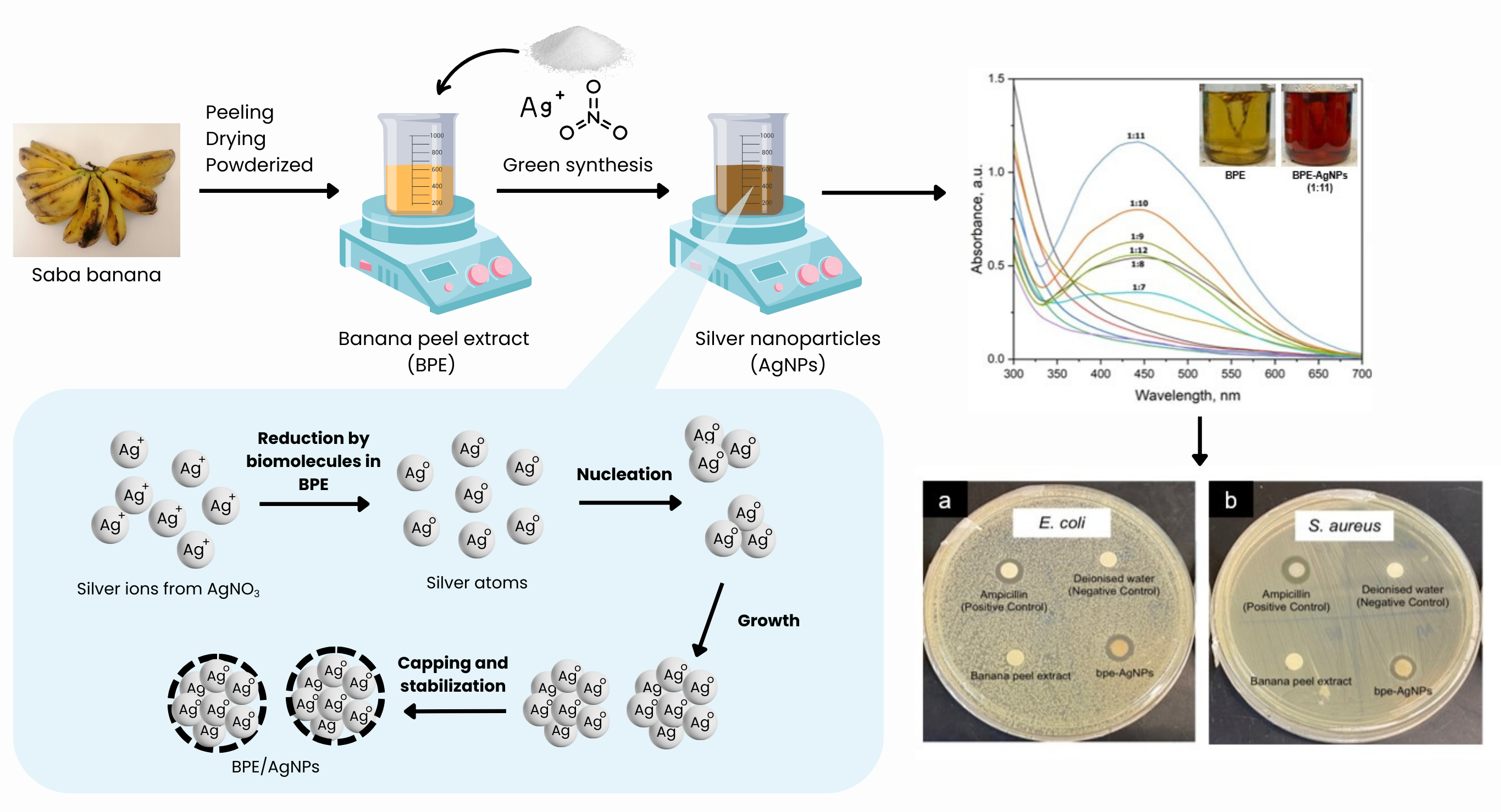
Synthesis of AgNPs was carried out using Saba BPE as green reducing agent and silver nitrate as metal salt precursor following protocol adapted from Jaffar et al. (2024) as illustrated in Figure 1. The effect of different volume ratios of BPE:AgNO3 on nanoparticle formation were evaluated in the range of 1:1 to 1:12. Specifically, 20 ml BPE aliquots were mixed with 20 to 240 ml of AgNO3 solution. The reaction mixtures were heated on a hot plate stirrer at 90°C for 2 h. The formation of AgNPs was indicated by a distinct color change from pale yellow to dark yellowish-brown due to the reduction of Ag⁺ to metallic Ag⁰. After synthesis, the mixtures were cooled to ambient temperature (25 °C) and subjected to centrifugation (7,500 × g, 5 min) to concentrate the nanoparticles. The resulting pellets were washed three times with ethanol to remove residual solvent and subsequently air-dried at room temperature for 24 h. The final biosynthesised nanoparticles (denoted as BPE-AgNPs) were stored in sterile amber vials at 4°C prior to characterization and antibacterial evaluation.
Figure 1. Processes involved in green synthesis of AgNPs from Saba banana peel extract.
Characterization of BPE-AgNPs
Synthesis of BPE-AgNPs were monitored using a UV-Visible spectrophotometer (Thermo Scientific NanoDrop 2000c). Deionised water was used as blank. The absorbance spectrum was recorded across the visible region of electromagnetic spectrum (400 – 700 nm). The presence of a distinct surface plasmon resonance (SPR) peak at this region indicates the reduction of Ag⁺ ions to metallic Ag⁰ and the formation of AgNPs. To investigate the chemical interactions between BPE and the synthesised AgNPs, FT-IR spectroscopy (Agilent Cary 630, Agilent Technologies Inc.) analysis was performed. Spectra were obtained in the range of 500–4000 cm⁻¹ at room temperature. The morphological information including particle size, shape and structure was obtained using Scanning Electron Microscope (Hitachi S3400N) and Transmission Electron Microscope (Tecnai G2 Spirit BioTWIN). Prior to analysis, the sample was mounted on a stub using double-sided tape. The sample was observed under vacuum at an accelerating voltage of 15 kV. It is worth mentioning that sputter coating was not required for this sample to eliminate disturbance from gold element during analysis. The elemental composition was determined using energy-dispersive X-ray spectroscopy (EDX) (Bruker Nano GmbH) connected to the SEM with the primary beam energy set at 15 keV.
Antibacterial Activity of BPE-AgNPs
Disk diffusion assay was performed to evaluate the antibacterial activity of BPE-AgNPs following the protocol adapted from Jaffar et al. (2024). Staphylococcus aureus and Escherichia coli as representative of Gram-positive and Gram-negative bacteria, respectively. Bacterial cells were retrieved from glycerol stocks, streaked onto the Luria-Bertani (LB) agar plates and incubated overnight at 37°C to obtain single colonies. Individual colonies were subsequently inoculated into LB broth and incubated overnight at 37°C to prepare bacterial cultures for antibacterial assays. Sterile solutions of ampicillin, BPE and BPE-AgNPs were prepared at a concentration of 1.0 mg/mL in a biological safety cabinet. Bacterial suspensions were spread evenly on Mueller-Hinton agar by using sterile swabs. After that, sterile blank discs (6 mm) were impregnated with 10 μl of the sample solutions, dried and placed onto the inoculated agar plates. Sterile water and ampicillin were used as the negative and positive control, respectively. The agar plates were incubated overnight at 37 °C, and inhibition zones were measured to assess antibacterial activity. All assays were performed in triplicate (n=3). Data were presented as mean ± standard deviation.
Results
Green Synthesis of AgNPs Using Different BPE:AgNO₃ Ratios
Silver nanoparticles were successfully synthesised using BPE as a green reducing and stabilizing agent. As shown in Figure 2, nanoparticles formation was initially indicated by a shift in color from light yellow to dark brown after heating the reaction mixture at 90 °C for 2 hours. The synthesised AgNPs were further evaluated using UV-Vis spectroscopy to monitor nanoparticle formation and determine the optimal BPE:AgNO3 ratio. The spectra of BPE and AgNO3 alone displayed no characteristic absorption peak, confirming the absence of surface plasmon resonance (SPR) associated with AgNPs formation. When BPE and AgNO3 were mixed, distinct SPR bands between 400 nm to 500 nm were observed, indicating the formation of AgNPs. At lower precursor concentrations (1:1 to 1:6), no SPR peaks were recorded. Increasing the AgNO₃ concentration (1:7 to 1:12), resulted in the appearance of characteristic SPR peak of AgNPs. The λmax values of the synthesized AgNPs ranged from 438 to 450 nm, with the highest wavelength recorded at the 1:8 ratio (450 nm). The λmax was then blue-shifted and stabilized at 443 nm for ratios between 1:9 and 1:12. The highest absorption intensity was recorded at a BPE: ratio of 1:11, while a subsequent decline in peak intensity was observed at the 1:12 ratio. Based on the UV–Vis spectral characteristics, the BPE:AgNO₃ ratio of 1:11 was selected as the optimum synthesis condition for subsequent characterization.
Figure 2. UV–Vis spectra of BPE, AgNO3 and BPE-AgNPs synthesised at different BPE:AgNO₃ ratios. Inset images show BPE before the reaction and BPE-AgNPs obtained after the reaction with AgNO₃ at a BPE:AgNO₃ ratio of 1:11.
Characterization of BPE-AgNPs
FTIR Spectroscopic Analysis
The FTIR spectrum of BPE-AgNPs is presented in Figure 3. Several absorption bands were observed within the range of 500–4000 cm⁻¹. A broad absorption band at 3671 cm⁻¹ was assigned to O–H stretching vibrations. Peaks at 2992 and 1465 cm⁻¹ corresponded to C–H stretching vibrations associated with aliphatic compounds. A minor peak at 2425 cm⁻¹ was attributed to carboxylic acid (-COOH) stretching vibrations. The absorption bands observed between 2000 and 2181 cm⁻¹ were assigned to C≡C stretching vibrations associated with alkyne or nitrile functional groups. A weak absorption band at 1819 cm⁻¹ corresponded to C=O stretching vibrations. Furthermore, peaks at 1139 cm⁻¹ and 837 cm⁻¹ were attributed to C–N stretching vibrations of aliphatic amines and aromatic C–H bending vibrations, respectively.
Figure 3. FT-IR spectra of BPE-AgNPs.
Morphology and Size Distribution Analysis
The morphology and size distribution of BPE-AgNPs were evaluated using SEM and TEM analysis (Figure 4). SEM imaging revealed a moderate degree of nanoparticle aggregation (Figure 4a). To obtain precise morphological information, TEM analysis was performed which revealed spherical nanoparticles with relatively inform distribution (Figure 4b). The average particle diameter determined from TEM analysis was 5.37 ± 1.36 nm (Figure 4c).
Figure 4. (a) SEM image of BPE-AgNPs; (b) TEM image of BPE-AgNPs; (c) Corresponding particle size distribution graph of the TEM image analyzed using ImageJ.
Elemental Composition Analysis
The elemental composition of BPE-AgNPs was analyzed using EDX spectroscopy (Figure 5). A strong silver signal above 3 keV was detected which confirmed the presence of metallic silver within the synthesised nanoparticles. Additional signals corresponding to carbon (C), oxygen (O), chlorine (Cl), and sulfur (S) were also observed. Quantitative EDX analysis revealed that silver represented the major component of the sample, accounting for 73.87 wt% of the sample composition, followed by chlorine (15.45 wt%), carbon (6.12 wt%), oxygen (3.63 wt%) and sulfur (0.94 wt%) (Table 1).
Figure 5. EDX spectra of BPE-AgNPs.
Table 1. Elemental composition of BPE-AgNPs
|
Elements |
Weight % |
Atomic % |
|
Carbon |
6.12 |
27.01 |
|
Oxygen |
3.63 |
12.03 |
|
Chlorine |
15.45 |
23.10 |
|
Silver |
73.87 |
36.31 |
|
Sulfur |
0.94 |
1.55 |
|
TOTAL |
100.00 |
100.00 |
Antibacterial Activity of BPE-AgNPs
The antibacterial activity of BPE-AgNPs against Escherichia coli and Staphylococcus aureus was evaluated using the disc diffusion assay (Figure 6). Clear inhibition zones were observed surrounding the discs containing BPE-AgNPs, whereas no inhibition zones were observed for the negative control (deionised water) and BPE alone. The diameter of zones of inhibition is summarized in Table 2. The BPE-AgNPs exhibited inhibition zones of 10.0 ± 0.0 mm against E. coli and 8.0 ± 0.0 mm against S. aureus. The positive control, ampicillin, produced inhibition zones of 13.3 ± 0.6 mm and 12.3 ± 0.6 mm against E. coli and S. aureus, respectively.
(a)
(b)
Figure 6. Representative agar plates from disk diffusion analysis of BPE-AgNPs (1.0 mg/mL) against the bacteria (a) E. coli and (b) S. aureus. Ampicillin and deionised water were used as positive and negative controls, respectively.
Table 2. Zone of inhibition from the disk diffusion assay.
|
Bacteria |
Zone of Inhibition (mm)* | |||
|
Deionised Water |
BPE |
Ampicillin (0.1 mg/mL) |
BPE-AgNPs (1.0 mg/mL) | |
|
E. coli |
0.0 ± 0.0 |
0.0 ± 0.0 |
13.3 ± 0.6 |
10.0 ± 0.0 |
|
S. aureus |
0.0 ± 0.0 |
0.0 ± 0.0 |
12.3 ± 0.6 |
8.0 ± 0.0 |
|
*Average value from triplicate measurements (n=3). | ||||
Discussion
Mechanism of BPE-Mediated AgNPs Formation
The successful formation of AgNPs was confirmed by the development of the brown coloration and the appearance of SPR bands in the UV–Vis spectra. The brown color formation after reaction between BPE and AgNO₃ is associated with collective oscillation of conduction electrons on the AgNPs surface following reduction of Ag⁺ ions into metallic Ag⁰ nanoparticles (Hosny et al., 2025). The absence of SPR signals from BPE and AgNO₃ alone indicates that nanoparticle formation occurred only after interaction between silver ions and phytochemical constituents present in BPE (Jaffar et al., 2023). The SPR response was strongly influenced by the BPE:AgNO₃ ratio, demonstrating the importance of precursor availability in controlling nucleation and nanoparticle growth (Tesfaye et al., 2023). At lower AgNO₃ concentrations (1:1–1:6), limited nanoparticle formation may have occurred due to insufficient silver ion availability for effective nucleation. Increasing AgNO₃ concentration enhanced nanoparticle formation, resulting in increased SPR intensity. However, excessive silver precursor concentration at the 1:12 ratio reduced the SPR intensity, suggesting decreased nanoparticle stability possibly due to uncontrolled particle growth or aggregation (Chouhan & Guleria, 2020; Savvidou et al., 2024) .
The optimum synthesis condition observed at a BPE:AgNO₃ ratio of 1:11 indicates that an appropriate balance between silver ion reduction and phytochemical-mediated stabilization was achieved. During green synthesis, biomolecules present in BPE function as electron donors, facilitating the conversion of Ag⁺ into Ag⁰, while simultaneously acting as stabilizing and capping agents to regulate nanoparticle growth. This dual functionality represents a major advantage of plant-mediated synthesis, as it eliminates the requirement for additional synthetic reducing and stabilizing chemicals.
The proposed mechanism of BPE-mediated AgNPs formation is illustrated in Figure 7. The phytochemical constituents in BPE initiate electron transfer to silver ions (Ag⁺), resulting in the formation of silver atoms (Ag°). These atoms subsequently undergo nucleation to form small nuclei. The growth of these nuclei continues as additional Ag⁺ ions are reduced and attached to the existing particles, leading to the formation of larger nanoparticles. The process continues until a dynamic equilibrium is achieved, where the rates of nucleation and growth offset each other. This mechanism emphasizes the dual role of BPE as both a reducing and stabilizing agent in the synthesis of AgNPs (Gün Gök et al., 2021; Thanikodi, 2026; Yi et al., 2022) .
Figure 7. Mechanism of BPE-AgNPs formation through reduction of Ag+ to Ag0 by biomolecules present in BPE followed by nucleation, growth, capping and stabilization.
Physicochemical Properties of BPE-AgNPs
The FTIR analysis provided evidence for the involvement of oxygen-, carbonyl-, and nitrogen-containing functional groups derived from banana peel phytochemicals in AgNPs formation. Negatively charged groups such as hydroxyl (O-H) and carbonyl (C=O) exhibit strong affinity for Ag⁺ ions due to their electron-donating capacity, thereby facilitating the reduction of Ag⁺ to Ag⁰ (Pirtarighat et al., 2019; Thongnopkun et al., 2018) . These findings are consistent with earlier studies which demonstrated that functional groups in plant extracts play a significant role in nanoparticle synthesis. Following reduction, biomolecules from BPE can adsorb onto the nanoparticle surface, forming an organic corona that prevents excessive aggregation and improves colloidal stability. Banana peel contains abundant cellulose, hemicellulose, pectin, phenolic compounds and other secondary metabolites, which collectively provide reducing and capping functions. Therefore, the phytochemical composition of BPE plays a critical role in controlling nanoparticle formation and maintaining colloidal stability.
The TEM analysis revealed spherical nanoparticles with an average diameter of 5.37 ± 1.36 nm, indicating efficient nucleation and growth regulation by BPE-derived biomolecules. Although slight aggregation is also observed, the nanoparticles remain spatially separated due to a thin layer of organic corona around the nanoparticles that imparts colloidal stability and biocompatibility. Ibrahim (2015) also reported spherical morphology for AgNPs derived from Musa paradisiaca BPE but with significantly larger size (23.7 nm) while some studies report spherical particles without clearly defined size distribution (Goh et al., 2023; Mishra, 2021) . This indicates that the BPE-mediated system in the present study yields comparatively smaller and more uniformly dispersed nanoparticles. Furthermore, the ultrasmall size of BPE-AgNPs is consistent with the previously reported biogenic AgNPs such as those synthesised using Solanum nigrum leaf extract (3.45 - 8.79 nm) (Abd-elhamed et al., 2025), Lysiloma acapulcensis extract (5 nm) (Garibo et al., 2020) and Bacillus subtilis (3 – 20 nm) (Alsamhary, 2020). The variation in particle size across different studies are probably influenced by factors such as differences in phytochemical composition, reduction kinetics and capping efficiency. The ultrasmall size of BPE-AgNPs provides a high surface area-to-volume ratio, which may enhance interactions with biological targets and contribute to their antibacterial activity.
Although SEM analysis showed some degree of aggregation, this phenomenon is commonly observed during nanoparticle characterization due to sample drying effects. During drying, water molecules migrate from the centre of the droplet towards the edge, carrying suspended nanoparticles and causing them to accumulate at the droplet perimeter. This process promotes localized aggregation in the absence of a dispersing medium (Goh et al., 2023; Jaffar et al., 2023). Nevertheless, TEM analysis confirmed that the nanoparticles remained individually distinguishable, suggesting that BPE-derived biomolecules effectively stabilized the nanoparticles and minimized irreversible aggregation. The EDX analysis further confirmed successful formation of metallic AgNPs, with silver representing the dominant elemental component. The absence of nitrogen originating from AgNO₃ suggests effective conversion of silver ions during synthesis (Molina et al., 2019). Meanwhile, residual carbon, oxygen, sulfur and chlorine signals are likely derived from the phytochemicals present in the BPE which form organic-inorganic mixed precipitates or residual capping agents on the nanoparticle surface (Ibrahim, 2015). Such biomolecular residues are commonly reported in green‑synthesised nanoparticles and contribute to colloidal stability by forming protective layers around the metallic core (Gün Gök et al., 2021; Riaz et al., 2022; Wan et al., 2021).
Antibacterial Performance and Potential Applications of BPE-AgNPs
The antibacterial activity of BPE-AgNPs against both Gram-negative (E. coli) and Gram-positive (S. aureus) bacteria demonstrates their functional potential as antimicrobial nanomaterials. Gram-negative bacteria such as E. coli have thinner peptidoglycan layers, rendering them more susceptible to AgNPs penetration and subsequent disruption. In contrast, the thicker peptidoglycan wall of Gram-positive S. aureus provides greater resistance against nanoparticle penetration (Arsène et al., 2021; Huq, 2020). Kokila et al. (2015) reported a similar observation for AgNPs produced using Cavendish banana peel extract with maximum zone of inhibition of 10.75 mm against E. coli and K. pneumoniae. Yi et al. (2022) observed a considerably lower zone of inhibition (4 ± 0.05 mm) against E. coli which may be attributed to the variations in phytochemical composition among different banana varieties as well as the differences in physicochemical properties of the synthesized nanoparticles including size, stability and surface functionalization. Furthermore, similar antibacterial trends have been reported for other biogenic AgNPs systems with zone on inhibition ranging from 7.0 to 11.0 mm for E. coli and 6.0 to 8.7 mm for S. aureus at AgNPs concentrations of 0.05 to 1.0 mg/mL (Jaffar et al. 2024). These findings indicate that the antibacterial performance observed in the present study is comparable to previously reported biogenic AgNPs systems.
AgNPs are widely recognized for their potent antimicrobial properties, which are significantly enhanced at the nanoscale due to their high surface area and unique physicochemical characteristics. Unlike conventional antibiotics such as ampicillin, AgNPs are less likely to induce bacterial resistance due to their multitarget mode of action, which poses a greater challenge for bacterial adaptation compared to antibiotics that act through specific mechanisms. Although the precise antibacterial mechanism of AgNPs is not fully understood, several plausible pathways have been proposed (Dakal et al., 2016; Ribeiro et al., 2020; Vanlalveni et al., 2021) . AgNPs can interact directly with bacterial cell membranes, causing structural damage and increased permeability. Once internalized, they may interfere with respiratory enzymes, impair ATP production and induce the generation of reactive oxygen species (ROS). These ROS can interact with DNA, where sulfur and phosphorus are key components, leading to genotoxic effects that disrupt DNA replication and repair processes. The cumulative impact of these events ultimately results in bacterial cell death. Therefore, these findings highlight the potent and multifaceted antibacterial activity of BPE‑AgNPs as effective biogenic nanomaterials for antimicrobial applications.
Besides their physicochemical properties and antibacterial performance, from a sustainability perspective, the use of banana peel extract for green synthesis of AgNPs offers strong potential within a circular economy framework. This approach enables valorization of abundant agro-industrial waste into high-value nanomaterials while eliminating the need for hazardous chemicals which is align with United Nations Sustainable Development Goals Responsible, specifically Responsible Consumption and Production (SDG 12). In terms of scalability, the simplicity of the synthesis process which involves mild reaction conditions in aqueous media could enabled large-scale production with minimal energy input. Therefore, the integration of banana peel-mediated AgNP synthesis into industrial processes offers a promising pathway toward sustainable nanomaterial production with potential applications in biomedical, environmental, food and energy sectors (Gong et al., 2024; Kurul et al., 2025) .
Conclusions
This study successfully demonstrated the green synthesis of AgNPs using Saba banana peel extract as an effective bioreducing and stabilizing agent. The optimized BPE:AgNO₃ ratio of 1:11 produced AgNPs with a surface plasmon resonance peak at 443 nm. FTIR confirmed the involvement of hydroxyl, carbonyl and amine groups in nanoparticle formation. SEM analysis revealed spherical AgNPs with sizes below 100 nm, while EDX confirmed a high silver content (73.87 wt%), supporting the efficiency of the biogenic synthesis. The synthesised BPE-AgNPs displayed strong antibacterial activity against both Gram-negative (E. coli) and Gram-positive (S. aureus) bacteria. These findings reinforce the viability of banana peel waste as a sustainable and low-cost precursor for nanoparticle production, in alignment with circular economy principles. The approach not only valorizes agricultural waste but also provides an environmentally friendly alternative to conventional synthesis. Moving forward, further studies involving advanced characterization such as XRD and DLS are required to further validate the structural properties and colloidal stability of the biosynthesized AgNPs, and in-depth toxicological assessments are necessary to ensure biocompatibility and safe integration of BPE-AgNPs in various applications including biomedical, environmental, food and energy sectors.
Declaration of Generative AI and AI-assisted Technologies in the Writing Process
During the preparation of this manuscript the author(s) used ChatGPT and Co-Pilot to assist with language editing during manuscript preparation. After using this tool/service, the author(s) reviewed and edited the content thoroughly and take(s) full responsibility for the content of the publication.
Acknowledgments
The authors would like to acknowledge the financial support from Universiti Malaysia Sabah through the UMSGreat Grant Scheme (GUG0550-1/2022).
Compliance with ethics guidelines
The authors declare they have no conflict of interest or financial conflicts to disclose.
This article contains no studies with human or animal subjects performed by the authors.
References
- Madhusudanan, Mukil; Zhang, Jian; Pandit, Santosh; Singh, Priyanka; Jeong, Geum-jae; Khan, Fazlurrahman et al. 2025. Green Synthesis of Silver Nanoparticles : A Review of Polymer and Antimicrobial Drug Combinations for Enhanced Antimicrobial Applications. 2400194. https://doi.org/10.1002/anbr.202400194
- Venkatesan, Jayachandran; Gupta, Pramod K; Eun, Seong; Won, Son; Gi, Hur; Seong, Hun. 2023. Silver-Based Hybrid Nanomaterials : Preparations , Biological , Biomedical , and Environmental Applications. Journal of Cluster Science. 34(1). https://doi.org/10.1007/s10876-021-02212-3
- Hosny, Shimaa; Gaber, Ghalia A; Ragab, Mona S; Ragheb, Mohamed A; Anter, Mena; Mohamed, Lamiaa Z. 2025. A Comprehensive Review of Silver Nanoparticles ( AgNPs ): Synthesis Strategies , Toxicity Concerns , Biomedical Applications , AI-Driven Advancements , Challenges , and Future Perspectives. Arabian Journal for Science and Engineering. https://doi.org/10.1007/s13369-025-10612-0
- Ahmed, Omnia; Sibuyi, Nicole Remaliah Samantha; Fadaka, Adewale Oluwaseun; Madiehe, Madimabe Abram; Maboza, Ernest; Meyer, Mervin et al. 2022. Plant Extract-Synthesized Silver Nanoparticles for Application in Dental Therapy. Pharmaceutics. 14(2). https://doi.org/10.3390/pharmaceutics14020380
- Ejaz, Umer; Afzal, Muhammad; Mazhar, Modasrah; Riaz, Muhammad; Ahmed, Naveed; Rizg, Waleed Y. et al. 2024. Characterization, Synthesis, and Biological Activities of Silver Nanoparticles Produced via Green Synthesis Method Using Thymus Vulgaris Aqueous Extract. International Journal of Nanomedicine. 19. https://doi.org/10.2147/IJN.S446017
- Chandhirasekar, K.; Thendralmanikandan, A.; Thangavelu, Pazhanivel; Nguyen, Ba Son; Nguyen, Trong Anh; Sivashanmugan, Kundan et al. 2021. Plant-extract-assisted green synthesis and its larvicidal activities of silver nanoparticles using leaf extract of Citrus medica, Tagetes lemmonii, and Tarenna asiatica. Materials Letters. 287. https://doi.org/10.1016/j.matlet.2020.129265
- Vallinayagam, Sugumari; Rajendran, Karthikeyan; Sekar, Vigneshkumar. 2021. Green synthesis and characterization of silver nanoparticles using Naringi crenulate leaf extract: Key challenges for anticancer activities. Journal of Molecular Structure. 1243(November). https://doi.org/10.1016/j.molstruc.2021.130829
- Gogoi, Bhaskarjyoti; Kumar, Rupesh; Upadhyay, Jnanendra; Borah, Debajit. 2020. Facile biogenic synthesis of silver nanoparticles (AgNPs) by Citrus grandis (L.) Osbeck fruit extract with excellent antimicrobial potential against plant pathogens. SN Applied Sciences. 2(10). https://doi.org/10.1007/s42452-020-03529-w
- Aktepe, Necmettin; Baran, Ayşe. 2021. Biosynthesis of AgNPs by extract from waste leaves of Citrullus lanatus sp. (watermelon); Characterization, antibacterial and antifungal effects. Progress in Nutrition. 23(3). https://doi.org/10.23751/pn.v23i3.11907
- Hasan, Wafiqah Latuapo; Sari, Retno; Hendradi, Esti. 2024. Green Synthesis of Antimicrobial Silver Nanoparticles using Green Tea Extract: The Role of Concentration and pH. Jurnal Sains Farmasi & Klinis. 11(1). https://doi.org/10.25077/jsfk.11.1.25-31.2024
- Thanh, Nguyen Chi; Pugazhendhi, Arivalagan; Chinnathambi, Arunachalam; Alharbi, Sulaiman Ali; Subramani, Baskaran; Brindhadevi, Kathirvel et al. 2022. Silver nanoparticles (AgNPs) fabricating potential of aqueous shoot extract of Aristolochia bracteolata and assessed their antioxidant efficiency. Environmental Research. 208(January). https://doi.org/10.1016/j.envres.2022.112683
- Onsang, Nordin; Pebrian, Darius; Anggraini, Fivi. 2023. Sustainability of Malaysian smallholder banana farming: an energy efficiency use-based audit. Agricultural Engineering International: CIGR Journal. 25(1).
- Flores-Contreras, Elda A.; González-González, Reyna Berenice; Pablo Pizaña-Aranda, José Juan; Parra-Arroyo, Lizeth; Rodríguez-Aguayo, Arath A.; Iñiguez-Moreno, Maricarmen et al. 2024. Agricultural waste as a sustainable source for nanoparticle synthesis and their antimicrobial properties for food preservation. Frontiers in Nanotechnology. 6(February). https://doi.org/10.3389/fnano.2024.1346069
- Mohd Jamil, Nurhidayah Azmirah; Jaffar, Syafiqah Syazwani; Saallah, Suryani; Misson, Mailin; Siddiquee, Shafiquzzaman; Roslan, Jumardi et al. 2022. Isolation of Cellulose Nanocrystals from Banana Peel Using One-Pot Microwave and Mild Oxidative Hydrolysis System. Nanomaterials. 12(19). https://doi.org/10.3390/nano12193537
- Abonyi, Matthew N.; Aniagor, Chukwunonso O.; Obi, Christopher C.; Nwadike, Emmanuel C.. 2024. Green synthesis and characterisation of silver nanocomposites and nanoparticles derived from banana leaves. International Journal of Green Economics. 18(4). https://doi.org/10.1504/IJGE.2024.142407
- Weiming, G.; Quanfeng, H.; Jianxia, S.; Dan, L.; Xuejuan, D.. 2023. Green synthesis of silver nanoparticles using banana flower extract, and their antibacterial activity. International Food Research Journal. 30(3). https://doi.org/10.47836/ifrj.30.3.06
- Goh, Hui Tung; Cheok, Choon Yoong; Yeap, Swee Pin. 2023. Green synthesis of silver nanoparticles using banana peel extract and application on banana preservation. Food Frontiers. 4(1). https://doi.org/10.1002/fft2.206
- Saallah, Suryani; Roslan, Jumardi; Zakaria, Nurul Nadjwa; Pindi, Wolyna; Siddiquee, Shafiquzzaman; Misson, Mailin et al. 2020. Isolation of nanocellulose from Saba’ (Musa acuminata x balbisiana) banana peel by one-pot oxidation-hydrolysis system. Advances in Agricultural and Food Research Journal. 1(1). https://doi.org/10.36877/aafrj.a0000096.
- Jaffar, Syafiqah Syazwani; Saallah, Suryani; Misson, Mailin; Siddique, Shafiquzzaman; Roslan, Jumardi; Lenggoro, Wuled. 2024. Green synthesis, characterization and antimicrobial efficacy of silver nanoparticles from Kappaphycus alvarezii extract. Research on Chemical Intermediates. 50(7). https://doi.org/10.1007/s11164-024-05298-2
- Jaffar, Syafiqah Syazwani; Saallah, Suryani; Misson, Mailin; Siddiquee, Shafiquzzaman; Roslan, Jumardi; Lenggoro, Wuled. 2023. Green Synthesis of Flower-Like Carrageenan-Silver Nanoparticles and Elucidation of Its Physicochemical and Antibacterial Properties. Molecules. 28(2). https://doi.org/10.3390/molecules28020907
- Tesfaye, Melakuu; Gonfa, Yodahe; Tadesse, Getachew; Temesgen, Tatek; Periyasamy, Selvakumar. 2023. Green synthesis of silver nanoparticles using Vernonia amygdalina plant extract and its antimicrobial activities. Heliyon. 9(6). https://doi.org/10.1016/j.heliyon.2023.e17356
- Savvidou, Maria G.; Kontari, Evgenia; Kalantzi, Styliani; Mamma, Diomi. 2024. Green Synthesis of Silver Nanoparticles Using the Cell-Free Supernatant of Haematococcus pluvialis Culture. Materials. 17(1). https://doi.org/10.3390/ma17010187
- Chouhan, Sonam; Guleria, Sanjay. 2020. Green synthesis of AgNPs using Cannabis sativa leaf extract: Characterization, antibacterial, anti-yeast and α-amylase inhibitory activity. Materials Science for Energy Technologies. 3(June). https://doi.org/10.1016/j.mset.2020.05.004
- Gün Gök, Zehra; Karayel, Mine; Yiğitoğlu, Mustafa. 2021. Synthesis of carrageenan coated silver nanoparticles by an easy green method and their characterization and antimicrobial activities. Research on Chemical Intermediates. 47(5). https://doi.org/10.1007/s11164-021-04399-6
- Yi, Cheong Wei; Amran, Syazwani Itri; Hussin, Huszalina; Zawawi, Nurliyana Ahmad. 2022. Materials in Life Sciences Silver Nanoparticles Synthesis using Banana Peels Extract and Its in Silico Evaluation for Antibacterial Activity against Escherichia coli. 1(1).
- Thanikodi, Sathish. 2026. Green Synthesis and Process Optimization of Zinc Oxide Nanoparticles Using Pseudomonas fluorescens : Taguchi – ANN-Based Evaluation of Physico-Chemical Parameters. 10(1).
- Thongnopkun, P.; Jamkratoke, M.; Jitkam, Y.. 2018. Green synthesis and characterization of silver nanoparticle using natural reducing sugar from cultivated banana peel. Journal of Physics: Conference Series. 1144(1). https://doi.org/10.1088/1742-6596/1144/1/012159
- Pirtarighat, Saba; Ghannadnia, Maryam; Baghshahi, Saeid. 2019. Green synthesis of silver nanoparticles using the plant extract of Salvia spinosa grown in vitro and their antibacterial activity assessment. Journal of Nanostructure in Chemistry. 9(1). https://doi.org/10.1007/s40097-018-0291-4
- Ibrahim, Haytham M.M.. 2015. Green synthesis and characterization of silver nanoparticles using banana peel extract and their antimicrobial activity against representative microorganisms. Journal of Radiation Research and Applied Sciences. 8(3). https://doi.org/10.1016/j.jrras.2015.01.007
- Mishra, S Moshahary P. 2021. Synthesis of silver nanoparticles ( AgNPs ) using culinary banana peel extract for the detection of melamine in milk. Journal of Food Science and Technology. 58(2). https://doi.org/10.1007/s13197-020-04791-x
- Abd-elhamed, Waled; Mohamed, Abeer A; Saad, Zakaria Hassan. 2025. Green synthesis of silver nanoparticles mediated by Solanum nigrum leaf extract and their antifungal activity against pine pathogens.
- Garibo, Diana; Borbón-Nuñez, Hugo A.; León, Jorge N.Díaz; García Mendoza, Ernesto; Estrada, Iván; Toledano-Magaña, Yanis et al. 2020. Green synthesis of silver nanoparticles using Lysiloma acapulcensis exhibit high-antimicrobial activity. Scientific Reports. 10(1). https://doi.org/10.1038/s41598-020-69606-7
- Alsamhary, Khawla Ibrahim. 2020. Eco-friendly synthesis of silver nanoparticles by Bacillus subtilis and their antibacterial activity. Saudi Journal of Biological Sciences. 27(8). https://doi.org/10.1016/j.sjbs.2020.04.026
- Molina, Gustavo A.; Esparza, Rodrigo; López-Miranda, J. Luis; Hernández-Martínez, Angel R.; España-Sánchez, Beatriz L.; Elizalde-Peña, Eduardo A. et al. 2019. Green synthesis of Ag nanoflowers using Kalanchoe Daigremontiana extract for enhanced photocatalytic and antibacterial activities. Colloids and Surfaces B: Biointerfaces. 180(January). https://doi.org/10.1016/j.colsurfb.2019.04.044
- Riaz, Muhammad; Sharafat, Uzma; Zahid, Nafeesa; Ismail, Muhammad; Park, Jeongwon; Ahmad, Bashir et al. 2022. Synthesis of Biogenic Silver Nanocatalyst and their Antibacterial and Organic Pollutants Reduction Ability. ACS Omega. 7(17). https://doi.org/10.1021/acsomega.1c07365
- Wan, Hong; Li, Chanyun; Mahmud, Sakil; Liu, Huihong. 2021. Kappa carrageenan reduced-stabilized colloidal silver nanoparticles for the degradation of toxic azo compounds. Colloids and Surfaces A: Physicochemical and Engineering Aspects. 616(May). https://doi.org/10.1016/j.colsurfa.2021.126325
- Huq, Md Amdadul. 2020. Green synthesis of silver nanoparticles using pseudoduganella eburnea MAHUQ-39 and their antimicrobial mechanisms investigation against drug resistant human pathogens. International Journal of Molecular Sciences. 21(4). https://doi.org/10.3390/ijms21041510
- Arsène, Mbarga M.J.; Podoprigora, I.; Davares, Anyutoulou K.L.; Razan, Marouf; Das, M. S.; Senyagin, A. N.. 2021. Antibacterial activity of grapefruit peel extracts and green-synthesized silver nanoparticles. Veterinary World. 14(5). https://doi.org/10.14202/vetworld.2021.1330-1341
- Geetha, T Kokila P S Ramesh D. 2015. Biosynthesis of silver nanoparticles from Cavendish banana peel extract and its antibacterial and free radical scavenging assay : a novel biological approach. Applied Nanoscience. https://doi.org/10.1007/s13204-015-0401-2
- Dakal, Tikam Chand; Kumar, Anu; Majumdar, Rita S.; Yadav, Vinod. 2016. Mechanistic basis of antimicrobial actions of silver nanoparticles. Frontiers in Microbiology. 7(NOV). https://doi.org/10.3389/fmicb.2016.01831
- Vanlalveni, Chhangte; Lallianrawna, Samuel; Biswas, Ayushi; Selvaraj, Manickam; Changmai, Bishwajit; Rokhum, Samuel Lalthazuala. 2021. Green synthesis of silver nanoparticles using plant extracts and their antimicrobial activities: a review of recent literature. RSC Advances. 11(5). https://doi.org/10.1039/d0ra09941d
- Ribeiro, Ana I.; Modic, Martina; Cvelbar, Uros; Dinescu, Gheorghe; Mitu, Bogdana; Nikiforov, Anton et al. 2020. Effect of dispersion solvent on the deposition of PVP-silver nanoparticles onto DBD plasma-treated polyamide 6,6 fabric and its antimicrobial efficiency. Nanomaterials. 10(4). https://doi.org/10.3390/nano10040607
- Gong, Xianyun; Jadhav, Nilesh D.; Lonikar, Vishal; Kulkarni, Anil N.; Zhang, Hongkun; Sankapal, Babasaheb R. et al. 2024. An overview of green synthesized silver nanoparticles towards bioactive antibacterial, antimicrobial and antifungal applications. Advances in Colloid and Interface Science. 323(November 2023). https://doi.org/10.1016/j.cis.2023.103053
- Kurul, Fatma; Doruk, Beyzanur; Topkaya, Seda Nur. 2025. Principles of green chemistry: building a sustainable future. Discover Chemistry. 2(1). https://doi.org/10.1007/s44371-025-00152-9
License
License
Copyright (c) 2026 by Authors

This work is licensed under a Creative Commons Attribution 4.0 International License.


